67-year-old male presenting with a 6-month history of right lower quadrant abdominal pain, diarrhea (4-6 watery stools/day, occasionally bloody), 10 lb weight loss, and fatigue. Symptoms worsened over the past month, with episodes of postprandial pain and nocturnal diarrhea. Former smoker with a 20-pack-year history, quit 10 years ago. No prior history of IBD. Positive for low-grade fever.

A 67-year-old male presents with a 6-month history of diarrhea and weight loss. An SBFT with a focus on the RLQ and terminal ileum shows irregular mucosa, lack of organized valvulae conniventes, strictures, and skip lesions, consistent with the segmental involvement characteristic of Crohn’s disease. The colon appears normal. Differential diagnosis includes infectious enteritis and ischemic enteritis.
Ashley Davidoff MD TheCommonVein.net 139824 (003SB).

A 67-year-old male presents with a 6-month history of diarrhea and weight loss. An SBFT with a focus on the RLQ and terminal ileum shows irregular mucosa, lack of organized valvulae conniventes, strictures, skip lesions, and a pseudodiverticulum, consistent with the segmental involvement characteristic of Crohn’s disease. The colon appears normal. Differential diagnosis includes infectious enteritis and ischemic enteritis.
Ashley Davidoff MD TheCommonVein.net 139825 (003SB).

A 67-year-old male presents with a 6-month history of diarrhea and weight loss. An SBFT with a focus on the RLQ and terminal ileum (magnified in b – red asterisks) and shows diffuse irregular narrowing, irregular mucosa, (maroon arrowheads) lack of organized valvulae conniventes, strictures, (yellow arrowheads) and a pseudodiverticulum (green arrowhead), with mild upstream dilatation of the normal bowel (pink asterisk). The constellation of findings are characteristic and consistent with Crohn’s ileitis. Differential diagnosis includes infectious enteritis and ischemic enteritis as less likely possibilities.
Ashley Davidoff MD TheCommonVein.net 139825 (003SB).

A 67-year-old male presents with a 6-month history of diarrhea and weight loss and a one month history of worsening symptoms.
A CT scan in the axial projection in the deep pelvis (magnified in b) shows diffuse wall thickening and luminal narrowing, irregularity of the mucosa and 2 focal regions of more significant narrowing with mild upstream dilatation of the normal ileum. There is mild congestion and induration of the subtending mesentery suggesting creeping fat. The visualised portion of the ascending colon contains liquified stool and has otherwise normal appearance. The constellation of findings are characteristic and consistent with acute Crohn’s ileitis. Differential diagnosis includes infectious enteritis and ischemic enteritis as less likely possibilities.
Ashley Davidoff MD TheCommonVein.net 139832c (003SB).

A 67-year-old male presents with a 6-month history of diarrhea and weight loss and a one month history of worsening symptoms.
A CT scan in the axial projection in the deep pelvis (magnified in b) shows diffuse wall thickening and luminal narrowing (red asterisk), irregularity of the mucosa (maroon arrowheads) and 2 focal regions of more significant narrowing (yellow arrowheads), with mild upstream dilatation of the normal ileum (pink asterisk). There is mild congestion and induration of the subtending mesentery (enclosed in white loop) suggesting creeping fat. The visualised portion of the ascending colon contains liquified stool and has otherwise normal appearance (white asterisk). The constellation of findings are characteristic and consistent with acute Crohn’s ileitis. Differential diagnosis includes infectious enteritis and ischemic enteritis as less likely possibilities.
Ashley Davidoff MD TheCommonVein.net 139832cL (003SB).

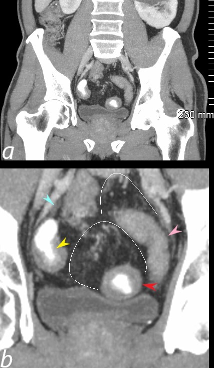
A 67-year-old male presents with a 6-month history of diarrhea and weight loss and a one month history of worsening symptoms. A CT scan in the coronal plane, with a focus on the pelvis (magnified in b), shows thickened loops of small bowel. The loop in the RLQ exhibits a thick wall with a narrowed lumen and alternating layers of enhanced and relatively non-enhanced wall. These stratifications are also appreciated as a “target sign” in the small bowel loop in the LLQ, which causes mass effect on the bladder. The loop abutting the left pelvic wall shows mild dilatation and dilution of contrast. Evidence of creeping fat in the mesentery subtending the affected loops is characterized by vascular and lymphatic congestion and mild induration. There is a small focus of loculated fluid along the right iliac vessels.
These findings are almost pathognomonic of acute Crohn’s ileitis. Included in the differential diagnosis, though less likely, are infectious enteritis and ischemic enteritis.
Ashley Davidoff MD TheCommonVein.net 139827c (003SB).

A 67-year-old male presents with a 6-month history of diarrhea and weight loss and a one month history of worsening symptoms. . A CT scan in the coronal plane, with a focus on the pelvis (magnified in b), shows thickened loops of small bowel. The loop in the RLQ exhibits a thick wall with a narrowed lumen and alternating layers of enhanced and relatively non-enhanced wall (yellow arrowhead) . These stratifications are also appreciated as a “target sign” in the small bowel loop in the LLQ, which causes mass effect on the bladder (red arrowhead) . The loop abutting the left pelvic wall shows mild dilatation and dilution of contrast (pink arrowhead). Evidence of creeping fat in the mesentery subtending the affected loops is characterized by vascular and lymphatic congestion and mild induration (enclosed in white ring). There is a small focus of loculated fluid along the right iliac vessels (teal arrowhead).
These findings are pathognomonic of acute Crohn’s ileitis. Included in the differential diagnosis, though less likely, are infectious enteritis and ischemic enteritis.
Ashley Davidoff MD TheCommonVein.net 139827cL (003SB).
References
- Magnetic Resonance Enterography for Predicting the Clinical Course of Crohn’s Disease Strictures. Schulberg JD, Wright EK, Holt BA, et al. Journal of Gastroenterology and Hepatology.
2.Creeping Fat Assessed by Small Bowel MRI Is Linked to Bowel Damage and Abdominal Surgery in Crohn’s Disease. Althoff P, Schmiegel W, Lang G, Nicolas V, Brechmann T. Digestive Diseases and Sciences. 2019;64(1):204-212.
3.The Mesenteric Fat and Intestinal Muscle Interface: Creeping Fat Influencing Stricture Formation in Crohn’s Disease. Mao R, Kurada S, Gordon IO, et al. Inflammatory Bowel Diseases. 2019;25(3):421-426.
Chaudhry NA, Riverso M, Grajo JR, et al. Inflammatory Bowel Diseases. 2017;23(4):641-649.
5.ACG Clinical Guideline: Management of Crohn’s Disease in Adults. Lichtenstein GR, Loftus EV, Isaacs KL, et al. The American Journal of Gastroenterology. 2018;113(4):481-517.
De Simone B, Davies J, Chouillard E, et al. World Journal of Emergency Surgery : WJES. 2021;16(1):23.
